Yue-Yang Zhang1,
Chang-Ting Huang2,
Shao-Ming Liu3 ![]() ,
Bin Wang4,
Jun Guo5,
Jian-Qi Bai6,
Xiao-Jing Fan7,
Yu-Sen Jia8
,
Bin Wang4,
Jun Guo5,
Jian-Qi Bai6,
Xiao-Jing Fan7,
Yu-Sen Jia8
For correspondence:- Shao-Ming Liu Email: liushaoming59@gmail.com Tel:+861052075325
Received: 15 July 2015 Accepted: 9 May 2016 Published: 28 June 2016
Citation: Zhang Y, Huang C, Liu S, Wang B, Guo J, Bai J, et al. Licochalcone A exerts antitumor activity in bladder cancer cell lines and mice models. Trop J Pharm Res 2016; 15(6):1151-1157 doi: 10.4314/tjpr.v15i6.6
© 2016 The authors.
This is an Open Access article that uses a funding model which does not charge readers or their institutions for access and distributed under the terms of the Creative Commons Attribution License (http://creativecommons.org/licenses/by/4.0) and the Budapest Open Access Initiative (http://www.budapestopenaccessinitiative.org/read), which permit unrestricted use, distribution, and reproduction in any medium, provided the original work is properly credited..
Purpose: To investigate the effect of licochalcone A (LA) on the inhibition of cell proliferation and ERK1/2 phosphorylation in bladder carcinoma cell lines.Methods: Cell viability was investigated using 3-(4,5-dimethylthiazol-2-yl)-2,5-diph-enyltetrazoliumbromide (MTT) assay. Dye-binding method was used to examine the concentration of proteins. Lymphocytes were extracted from mice and after surface staining were subjected to BD fixation and permeabilization for
intracellular staining. Flow cytometry was used to measure cellular fluorescence.
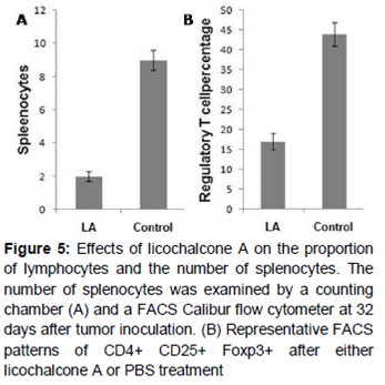
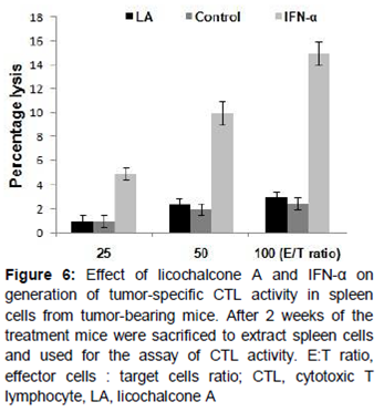
Results: MTT results revealed a significant decrease in the proliferation of UM-UC-3, J82 and HT-1197 cell lines on treatment with LA. LA also induced reduction in phosphorylation of ERK1/2 in all three carcinoma cell lines. In the mouse model, licochalcone A treatment via intraperitoneal (ip) administration induced a significant decrease in the level of regulatory T cells (Tregs). Comparison of the mouse interferon-α (IFN-α)-treated and LA-treated groups revealed that LA treatment caused enhancement of cytotoxic T lymphocyte (CTL) activity similar to that of IFN-α. Administration of UM-UC-3 cells in C3H/HeN mice resulted in marked reduction in the counts for splenocytes and CD4+ CD25+ Foxp3+ T (regulatory T cells) cell proportion in LA-treated mice compared to untreated control group.
Conclusion: Licochalcone A may be of therapeutic importance for the prevention of bladder carcinoma. However, studies are required to ascertain the compound’s usefulness in this regard.
Introduction
Cancer is the leading cause of death in economically developed countries and second leading cause of death in developing countries [1]. The tendency of cancer cells to proliferate uncontrollably, escape apoptosis and spread to other tissues makes it the most intractable disease [2]. Bladder carcinoma constitutes the most frequently detected cancer in industrialized countries [3]. It is associated with complex genetic events and may involve various molecular pathways, including the Ras-MAPK pathway [4]. Earlier reports show that RAS proteins transfer information from the receptors in cell surface to protein through different pathways [5]. It has been estimated that in 30 % of cancers RAS-RAF/MEK (mitogen extracellular kinase)/ERK (extracellular signal-related kinase) pathway is enhanced [6]. The first line of treatment for advanced renal cancers is sunitinib [7]. It is believed that suppression of tyrosine kinase can be a promising strategy for the inhibition of cancer cell proliferation. Tyrosine kinases mediate the binding of growth factors with their corresponding receptors [8].
Chalcones because of their potent biological activities have been the target of interest for the chemists as well as clinicians globally. They exhibit anti-inflammatory [9], anti-invasive [10], anti-tumour [11] and antibacterial [12] properties. Chalcone treatment causes induction of apoptosis and alters mitochondrial membrane potential in various types of cancers [13]. Licochalcone A (LA), an oxygenated chalcone was isolated from the roots of the Chinese liquorice (Glycyrrhiza uralensis). It is reported to exhibit various bioactive properties including anti-parasitic, estrogenic, antimalarial and antitumor activities [14]. LA regulates cell growth and induces cell apoptosis in prostate cancer cell lines [15]. In the present study the effect of licochalcone A (LA) on bladder carcinoma in human bladder cancer cells and mice model was investigated. The results revealed that licochalcone A treatment inhibits bladder cancer cell proliferation and reduces ERK1/2 phosphorylation. Furthermore, licochalcone A treatment caused a marked reduction in the regulatory T cell count in the mouse model. Also the cytotoxic T lymphocyte (CTL) activity was induced similar to that of IFN-α.
Methods
Drug and chemicals
Licochalcone A (LA) was isolated from the roots of the Chinese liquorice (Glycyrrhiza uralensis). The drug was dissolved in DMSO and stored at -20 ˚C. Mouse interferon-α (IFN-α) was purchased from BD Pharmingen (San Diego, CA). The antibodies against ERK1/2 (Thr202/Tyr204) and β-actin were purchased from Santa Cruz Biotechnology (Santa Cruz, CA).
Animals and ethical statement
Female C3H/HeN mice (12 weeks old) and female nu/nu mice (10 weeks old) were obtained from the Laboratory Animal Center, Third Military Medical University (Chongqing, China). The study was approved by the Ethics Committee of the Wangjing Hospital of China Academy of Chinese Medical Sciences (approval ref no. 201323765757/2014). All experimental animal procedures were conducted in compliance with State and Federal laws, standards of the US Department of Health and Human Services, and guidelines established by Tulane University Animal Care and Use Committee, accredited by Association for the Assessment and Accreditation of Laboratory Animal Care [16].
Tumor cell line
The bladder cancer cell lines, UM-UC-3, J82 and HT-1197 were obtained from The Cell Bank of Type Culture Collection of Chinese Academy of Sciences, Shanghai Institute of Cell Biology (Shanghai, China). The cells were cultured in DMEM containing antibiotics and were maintained in an incubator at 37 ˚C with humidified atmosphere of 5 % CO2.
MTT assay
Into a 96-well flat bottom micro plate (BD Falcon, Franklin, NJ) cells at a density of 2.5 x 10⁵ cells per well were distributed. To each of the well different concentration of licochalcone A was added and incubated for 72 h. MTT solution (10 μL) was added to each well and the plates were incubated for 4 h at 37 ˚C in 5 % CO2 incubator. Formazan crystals were dissolved by adding 0.04 N HCl in 2-propanol (100 μL). The microplate reader (MPR-A4i; Tosoh Corporation, Tokyo, Japan) was used to measure absorption at 570 nm. All the measurements were carried out in triplicate.
Western blotting
The cells treated with licochalcone A were lysed and the concentration of proteins was determined by dye-binding method (Bio-Rad). The proteins were resolved on 15 % SDS-PAGE and transferred to nitrocellulose membranes. Digitonin-based subcellular fractionation technique was used for cytosolic and mitochondrial fractions. Onto DS-PAGE equal volumes of cytosolic and mitochondrial fractions were resolved and transferred to nitrocellulose membranes. The membranes after incubation with primary antibody were washed, and then incubated with horseradish peroxidase anti-mouse or horseradish peroxidase antirabbit antibodies. Enhanced chemiluminescence system was used for visualization of immunoreactive bands.
Intracellular FACS
The lymphocytes extracted from the mice after surface staining were subjected to BD fixation and permeabilization (BD Biosciences) for intracellular staining using the manual protocol. After incubation with 100 μL of BD Cytofix/Cytoperm solution the cells were washed with BD perm-wash buffer and stained with FITC-conjugated anti-Foxp3 mAb (FJK-16s). FACS Calibur flow cytometer (BD Biosciences, San Jose, CA) was used to measure the cellular fluorescence.
Treatment strategy
UM-UC-3 cells at a density of 2.5 x 10⁷ were administered subcutaneously into the lateral flanks of the mice. When tumor achieved palpable stage, the mice in the treatment and control group were given licochalcone A (40 mg/kg) or phosphate buffered saline (PBS), respectively through the intravenous route for 4 weeks. After every 2 days the size of the tumor was measured with calipers and converted to volume.
Determination of CTL activity
The mice administered UM-UC-3 cells were sacrificed on day 14 following treatment with licochalcone A or PBS. The splenocytes were extracted and cultured with mitomycin C (MMC)-treated UM-UC-3 cells for 5 days after adding 25 ng/mL IL-2 (Takeda Chemical Industries, Tokyo, Japan). The Percoll (Amersham Biosciences, Piscataway, NJ) density gradient centrifugation was used to separate the viable cells whereas the effector cells were incubated with 51Cr-labeled UM-UC-3 cells for 6 h. After incubation the cytotoxicity was evaluated.
Statistical analysis
All the experiments were performed in triplicates and Student's t test was used for the determination of statistical significance. Differences were considered statistically significant at p < 0.05. Graph Pad Prism 5 (Graph Pad Software, Inc., San Diego, CA, USA) was used for the analysis of the data.
Results
Effect of licochalcone A on bladder cancer cell growth and ERK1/2 expression
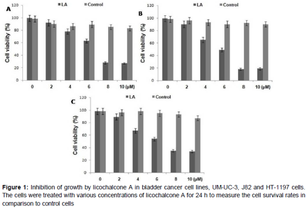
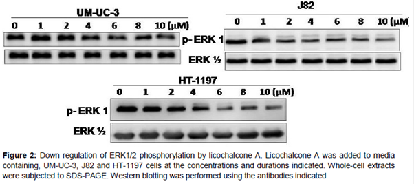
The effect of licochalcone A on the growth of UM-UC-3, J82 and HT-1197 bladder cancer cells was analyzed. It was observed that licochalcone A suppressed the growth of all the three tested bladder cancer cell lines in a dose-dependent manner (). Licochalcone A treatment also caused decrease in the phosphorylation of ERK1/2 in all the three tested bladder carcinoma cell lines (). ERK 1/2 has been reported to be involved in drug resistance [11-17].
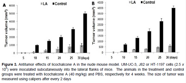
Antitumor effect of licochalcone A in nude mouse model
UM-UC-3, J82 and HT-1197 at a density of 2.5 x 10⁶ was administered to each of the mice. After the tumor attained palpable stage, the animal in the treatment and control groups were given licochalcone A (40 mg/kg) and saline, respectively. Analysis of the animals revealed that tumor volumes in control group were markedly higher compared to treatment group ().
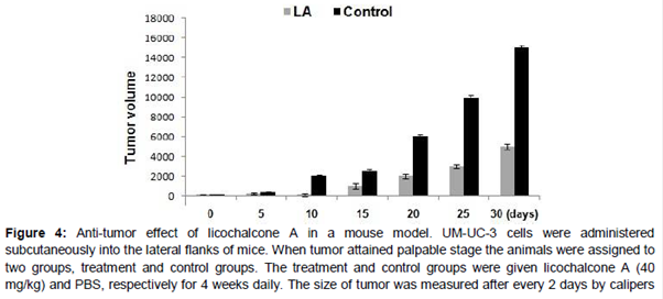
Antitumor effect of licochalcone A in the C3H/HeN mouse model
The mice were treated with 40 mg/kg doses of the licochalcone A on the day 4 after administration of 2.5 x 10⁵ UM-UC-3 cells. Tumor growth was significantly (p < 0.005) reduced in the licochalcone A treated mice compared with those in the control group ().
Effect of licochalcone A on T cells and generation of tumor-specific CTL activity in splenocytes of tumor-bearing C3H/HeN mice
The effect of licochalcone A was also examined on effector cells for anti-tumor activity using flow cytometry on day 21 after administration of UM-UC-3cells in C3H/HeN mice. The results showed a marked reduction in the counts for splenocytes and CD4+ CD25+ Foxp3+ T (regulatory T cells) cell proportion in the licochalcone A treated mice compared to untreated control group (A, B).
The effect of licochalcone A on the level of CTL activity was also investigated and compared with IFN-α-treated mouse. It was observed that the level of CTL activity was significantly higher in IFN-α-treated mouse compared to licochalcone A treated mouse ().
Discussion
Bladder carcinoma constitutes the most frequently detected cancer in industrialized countries and is associated with complex genetic events involving various molecular pathways, including the Ras-MAPK pathway [3,4]. In the present study use of licochalcone A in bladder carcinoma treatment was investigated. The results revealed that licochalcone A significantly suppressed the proliferation of bladder carcinoma cells and inhibited the tumor growth in mouse models. It is well known that ERK1/2 belonging to MAPK pathway plays a crucial role for the growth and survival of carcinoma cells [17]. It also induces resistance to various drugs used against cancers [18,19]. Therefore, it is believed that inhibition of ERK1/2 signaling pathway can be of great therapeutic importance for cancer treatment. The results from the present study showed that licochalcone A treatment inhibited phosphorylation of ERK1/2.
In patients suffering from chronic myelogenous leukemia the CTL activity is promoted on treatment with IFN-α [20,21]. The results of the present study revealed that licochalcone A treatment proved more effective for the improvement of CTL activity in the spleen compared to IFN-α. The CD4+CD25+Foxp3+ Tregs cell count in the licochalcone A treated group was significantly decreased compared to untreated group. Tregs have been shown to produce immunosuppressive cytokines as well as negative regulation of immunity [22]. Therefore, reduction in Tregs will promote immunity and enhance activity of the T-cells. In the licochalcone A treated mice the activity of CTL was significantly increased than those of untreated control mice. In addition the splenocyte count was also decreased in licochalcone A treated mice.
Conclusion
The results obtained show that licochalcone A has an anti-tumor effect in murine models of bladder cancer when used as a single agent. Furthermore, the findings suggest that licochalcone may enhance the therapeutic efficacy of existing immune-based therapies for metastatic cancer patients.
References
Archives


News Updates